
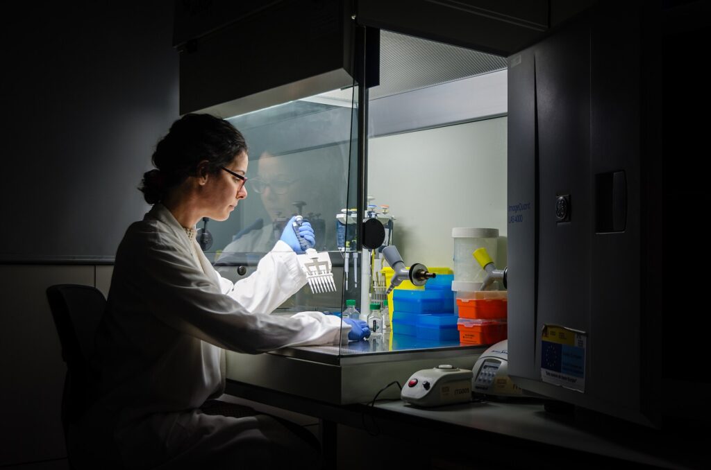
A recent study has demonstrated that modifying magnetic nanoparticles can significantly improve the isolation of RNA from biological samples—particularly when RNA is present at very low concentrations. This advancement builds on the growing need for reliable, rapid diagnostic tools in medical research and clinical settings, especially in the context of infectious diseases like COVID-19.
What Happened: A Breakthrough in RNA Isolation
Researchers have developed a method that uses specially modified magnetic nanoparticles to extract RNA more efficiently than traditional techniques. The original source, published on Phys.org, describes how these nanoparticles are engineered to bind specifically to RNA molecules in complex biological matrices such as blood, saliva, or tissue samples.
When exposed to a magnetic field, the particles can be easily separated from the sample after binding, allowing for clean recovery of RNA. This process reduces contamination and loss of RNA, which is a common issue in conventional isolation methods. The technique has been tested in laboratory settings with samples containing low levels of RNA, where it outperformed standard protocols in both yield and purity.
Key Facts from the Study
- The magnetic nanoparticles are chemically modified to enhance their affinity for RNA strands.
- After binding, the particles can be rapidly separated using a magnetic field, enabling quick and clean recovery of RNA.
- The method has shown improved performance in samples with low RNA concentrations—critical for early disease detection or monitoring.
- It is particularly useful for applications requiring small sample volumes, such as point-of-care diagnostics or personalized medicine.
- While the study is still in the laboratory phase, it represents a scalable and repeatable approach to nucleic acid isolation.
How It Works: A Step-by-Step Process
The process begins with the introduction of magnetic nanoparticles into a biological sample. These nanoparticles are functionalized with ligands—molecular structures that selectively bind to RNA—ensuring that only RNA molecules are captured.
After incubation, the sample is subjected to a magnetic field. This pulls the nanoparticle-RNA complexes out of the liquid phase, leaving behind unbound components. The recovered complexes are then washed and treated to release the RNA in a purified form, ready for downstream applications such as sequencing or gene expression analysis.
Unlike traditional methods that rely on chemical reagents or column-based filtration, this approach avoids harsh solvents and reduces the risk of RNA degradation. The magnetic separation step also enables automation, making it suitable for integration into high-throughput diagnostic platforms.
Why It Matters: Implications for Diagnostics and Research
RNA is a vital biomarker in disease detection and monitoring. For instance, viral RNA in respiratory samples is used to diagnose infections like influenza or SARS-CoV-2. However, RNA is inherently unstable and degrades quickly in biological fluids, especially at low concentrations.
Improved RNA isolation methods directly impact the sensitivity and reliability of diagnostic tests. With this new technique, researchers can detect RNA at lower levels, enabling earlier diagnosis and more accurate monitoring of disease progression. This is especially valuable in rare or early-stage conditions where RNA levels are minimal.
Additionally, the method supports applications in personalized medicine, where RNA profiles can inform treatment decisions. It also opens doors for point-of-care testing—where diagnostics are performed outside traditional labs—by reducing the need for complex equipment and lengthy processing times.
For example, a similar technology was recently used in a laser-based test that detects colorectal cancer biomarkers in blood in minutes, highlighting the broader trend toward rapid, accessible diagnostics. This new RNA isolation method could serve as a foundational tool in such systems.
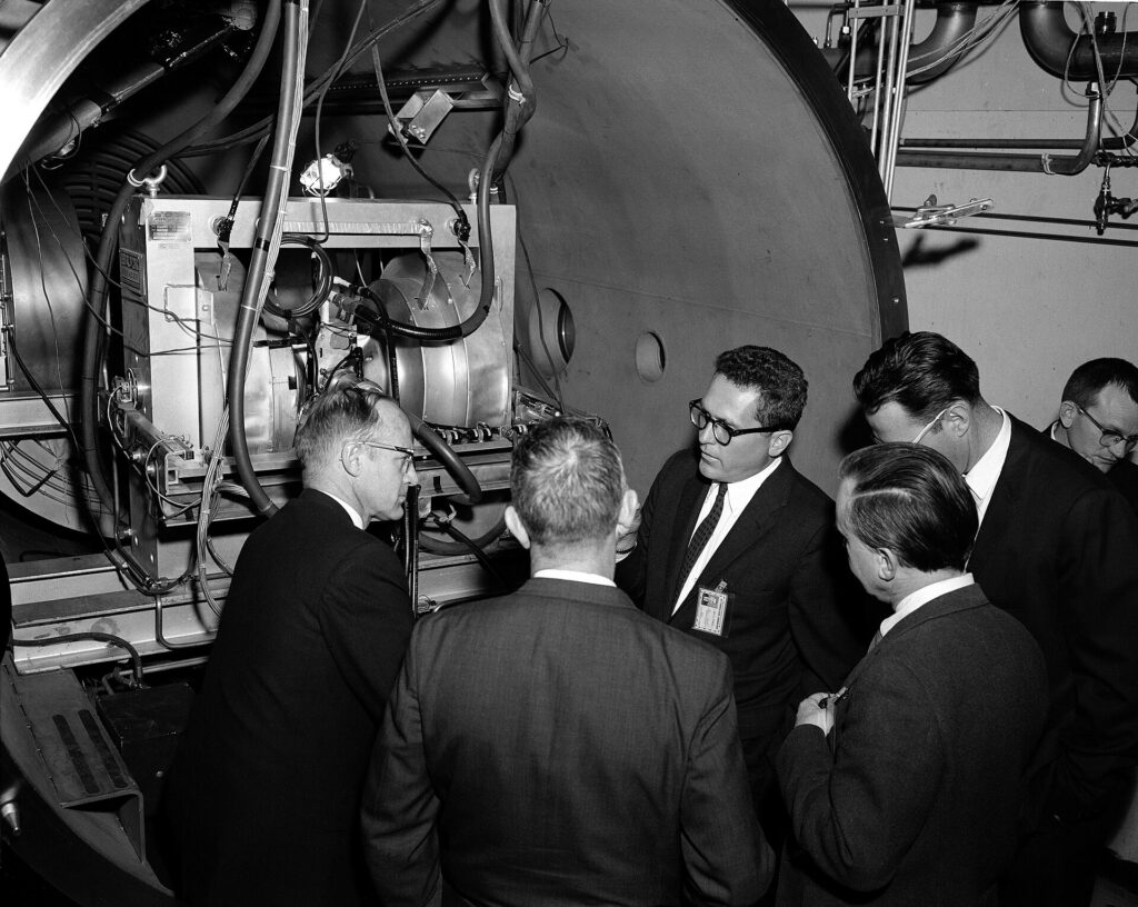
Lab-Oratory, the JPL employee newspaper, covered the opening of this new facility, describing how plasma can be generated by bodies entering an atmosphere at high speed and in the plasma lab by electrical discharge. The plasma facility at JPL could create thermally ionized gases at temperatures up to 30,000 degrees Fahrenheit. Findings from the plasma program were to be applied to power and propulsion devices, and Earth re-entry problems (thermal protection, communication blackout and electrical breakdown). This was a $1.6 million JPL task – part of the larger NASA plasma research and development program.
Plasma demonstration attended by Dr. William Pickering, Mr. Sparks, and Goddard.
March 4, 1964
Requested by: Collela
Photo by: J. Rayle by NASA, Public domain, via Wikimedia Commons. · Source
Limitations and Open Questions
While promising, the study has several limitations. The research is currently confined to laboratory-scale experiments and has not yet been validated in real-world clinical settings. The long-term stability of the modified nanoparticles and their potential toxicity in human tissues remain untested.
Additionally, the method’s performance across diverse biological matrices—such as different types of blood, urine, or tissue—has not been fully evaluated. Variability in sample composition could affect binding efficiency and RNA recovery.
Another open question is scalability. While the magnetic separation is efficient, the cost and availability of high-purity, functionalized nanoparticles may limit widespread adoption, especially in resource-limited environments.
What to Watch Next
Future research will likely focus on optimizing nanoparticle design for broader sample compatibility and enhanced stability. Scientists may explore combining this method with other technologies—such as microfluidic devices or AI-driven analysis—to create integrated diagnostic platforms.
Further validation in clinical trials will be essential to determine whether this method can reliably detect diseases in real-world samples. Researchers may also investigate how the technique performs in combination with other biomarkers or in the presence of inhibitors like proteins or lipids.
As this technology matures, it could become a standard component in diagnostic pipelines. For instance, the same principles used here could be adapted for isolating other nucleic acids, such as DNA or miRNA, expanding its utility across biomedical fields.
For readers interested in related developments, consider exploring: how nanoparticles are being used to restore immune function in cancer models, or how buffer conditions affect mRNA stability in delivery systems. These studies illustrate the broader role of nanoparticles in biotechnology.
Additionally, a laser-based test that detects colorectal cancer biomarkers in blood in minutes demonstrates the trend toward rapid, non-invasive diagnostics—where improved sample preparation is a key enabler.
Meanwhile, research into sparse silver nanoparticle layers enhancing CO₂-to-CO conversion shows how nanomaterials can be engineered for specific chemical transformations—another example of how surface modification drives functional performance.
Sources & further reading
Featured image: Researcher at work in her laboratory. Working every day until late is almost a common routine for a researcher that really believe in science progress. In this picture, PhD Mileidys Perez working at Vall d'Hebron Institute of Research (VHIR), Barcelona, Spain. by axventura, CC BY 4.0, via Wikimedia Commons. Image source · License